New Patients
Existing Patients

New Patients
Existing Patients

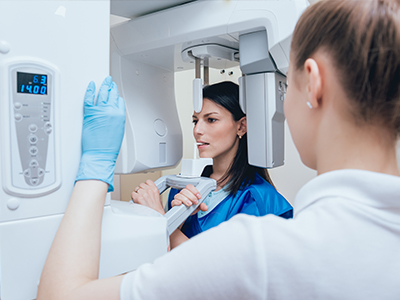
Digital radiography replaces traditional film with high‑sensitivity electronic sensors and computer processing to capture dental images. Instead of developing film in a darkroom, the image appears almost instantly on a monitor, allowing the dental team to review results in real time. This shift from chemical-based processing to digital acquisition has reshaped how dentists evaluate teeth, gums, and supporting bone.
For patients, the most noticeable change is the speed and clarity of the process. Images are captured quickly and can be enlarged, adjusted for contrast, and annotated without degrading the original file. That responsiveness helps clinicians explain findings more clearly during appointments and lets patients see the same images their provider uses to make decisions.
While the technology may sound technical, the experience in the chair is straightforward: sensors are positioned like film was in the past, and exposures are brief. The transition to digital hasn’t changed the fundamentals of safe radiographic practice; it has made the workflow faster and the diagnostic output easier to use for both clinicians and patients.
One of the primary advantages of digital radiography is lower radiation exposure compared with many conventional film methods. Because digital sensors require less radiation to produce a usable image, clinicians can obtain high‑quality diagnostic images with fewer x‑ray units. This reduction in dose contributes to safer routine care while still delivering the detail needed for accurate diagnosis.
Safety in dental imaging is achieved through a combination of technology and practice. Digital systems are used alongside established protection measures — for example, properly positioned sensors, modern x‑ray generators, and adherence to exposure guidelines — to minimize risk. When needed, clinicians will also use lead aprons or thyroid collars in accordance with current best practices.
Reducing radiation is just one part of a broader commitment to safety. Digital radiography also decreases the environmental hazards associated with chemical film processing, eliminating the need for development solutions and film waste that require special handling.
Digital images offer fine detail and flexibility that can improve diagnostic confidence. Clinicians can zoom in to inspect tiny fractures, evaluate the interface between a restoration and tooth structure, and assess bone levels around teeth and implants. Subtle findings that might be missed on poorly developed film are often more apparent on a manipulated digital image.
Software tools enhance this capability: clinicians can adjust brightness, contrast, and magnification; apply measurement overlays; and compare sequential images side by side. These features support everything from routine cavity detection to monitoring healing after periodontal therapy or implant placement.
Importantly, digital radiography complements — rather than replaces — a thorough clinical exam. Images are a powerful diagnostic aid, but they are interpreted in the context of an intraoral exam, patient history, and other diagnostic information to arrive at a complete treatment plan.
Digital files transform how dental records are managed. Once an image is taken, it is stored directly in the patient’s electronic chart where it can be retrieved instantly during follow‑up visits. This eliminates the need to file and locate physical film, streamlining recordkeeping and allowing staff to focus on patient care rather than administrative tasks.
Sharing images with other specialists or referring offices is also more efficient. Digital files can be sent securely and quickly, facilitating interdisciplinary collaboration when a case requires input from an oral surgeon, periodontist, or orthodontist. Faster information exchange reduces wait times in treatment planning and keeps communication clear and complete.
Longitudinal records are easier to maintain with digital imaging. Side‑by‑side comparisons across months or years support monitoring of progressive conditions and help clinicians identify changes earlier than might be possible with isolated film images.
When digital radiography is part of your visit, the process will feel familiar but faster. A small sensor is placed in or near the mouth for intraoral images, or an external scanner is used for panoramic views. Exposures are brief, and the resulting images appear on the operatory screen within seconds. Your clinician will review those images with you, pointing out areas of interest and explaining how they inform diagnosis and treatment options.
Because images are captured and stored digitally, your dentist can show you a clear visual representation of findings while discussing next steps. This shared view supports informed decision‑making and helps patients understand the rationale behind recommended care. If a specialist’s opinion is helpful, images can be shared promptly to ensure continuity of care.
At the office of Joanna Tricorache, DDS, digital radiography is one of several modern tools used to provide thorough, patient‑centered dental care at our Staten Island and Manhattan locations. We use imaging selectively and responsibly — only when it will provide information that affects diagnosis or treatment — and we follow accepted protocols to keep exposures as low as reasonably achievable.
In summary, digital radiography streamlines imaging, enhances diagnostic capability, and reduces radiation exposure while supporting clear communication between clinicians and patients. If you have questions about how we use digital imaging or whether it’s appropriate for your care, please contact us for more information.

Ready to schedule your next dental appointment or have questions about our services?
Contacting Joanna Tricorache, DDS is easy! Our friendly staff is available to assist you with scheduling appointments, answering inquiries about treatment options, and addressing any concerns you may have. Whether you prefer to give us a call, send us an email, or fill out our convenient online contact form, we're here to help. Don't wait to take the first step towards achieving the smile of your dreams – reach out to us today and discover the difference personalized dental care can make.