New Patients
Existing Patients

New Patients
Existing Patients

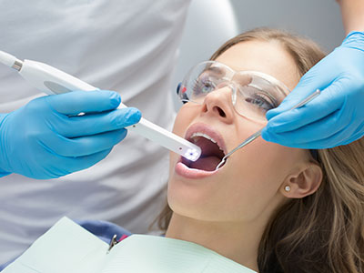
An intraoral camera is a small, pen-sized imaging device designed to capture high-resolution color images from inside the mouth. Unlike a traditional mirror exam, this tool provides close-up views of individual teeth, gums, and other oral tissues, allowing clinicians and patients to see the oral condition in real time. The images are displayed on a monitor, giving a magnified perspective that reveals details that might be hard to spot with the naked eye.
Because the camera produces clear, full-color images, it becomes an objective record of a patient’s oral health at a particular moment in time. These images complement visual inspection and radiographs by documenting surface conditions, restorative margins, and subtle soft-tissue changes. When used routinely, intraoral photography contributes to more informed decision-making and a higher standard of clinical documentation.
At our office, an intraoral camera is treated as a diagnostic and communication tool rather than a novelty. The device supports a careful, evidence-based approach to dental care: clinicians can confirm findings, cross-check results with other diagnostic tests, and explain recommended procedures with visual clarity. For patients, the result is more transparent care and a clearer understanding of oral health priorities.
One of the most practical benefits of intraoral cameras is that they make the invisible visible. When clinicians share live images with patients, it removes ambiguity from discussions about treatment needs. Seeing a high-resolution photo of a cracked tooth, a worn restoration, or inflamed gum tissue helps patients recognize the issue and better understand why a particular treatment may be recommended.
This visual approach changes the dynamic of the exam. Instead of relying solely on verbal descriptions, clinicians can walk patients through the anatomy and pathology on-screen, pointing out features and comparing them to healthy examples. That shared view builds trust and supports informed consent because patients are shown the same evidence their clinician uses to make recommendations.
For practices that emphasize patient education, the intraoral camera is a natural fit. It helps clinicians demonstrate proper hygiene challenges, track progress after interventions, and set realistic expectations for outcomes. As a result, patients who see their condition first-hand are often more engaged in preventive care and more comfortable with the clinical pathway proposed by the care team.
Intraoral cameras are versatile across many aspects of general and restorative dentistry. They excel at revealing surface-level defects such as hairline cracks, recurrent decay at margins, failing restorations, and early signs of erosion. Because they capture color and texture, cameras can also assist in identifying soft-tissue abnormalities that warrant observation or referral.
These cameras are particularly useful when combined with other diagnostic tools. A camera image can corroborate findings from a clinical exam or digital radiograph, giving clinicians a multi-modal perspective before deciding on treatment. In restorative planning, clear images of preparation margins and existing restorations guide laboratory communication and improve the fit and aesthetics of final prostheses.
In periodontal care, intraoral images document gingival inflammation, recession, and plaque accumulation in a way that is easy to archive and review. Over time, a series of images helps clinicians monitor healing after periodontal therapy, evaluate tissue response, and adjust maintenance protocols based on photographic evidence rather than memory alone.
Modern intraoral cameras integrate smoothly with electronic health records and imaging systems, enabling clinicians to attach time-stamped photos directly to a patient’s chart. This creates a searchable visual history that supports continuity of care and simplifies case reviews. Digital archiving also reduces the risk of losing important diagnostic data compared with paper-based notes or isolated files.
When collaboration is needed, intraoral images can be shared securely with specialists or dental laboratories to clarify clinical observations and treatment goals. A well-documented photo often eliminates ambiguity in referral communications and can streamline restorative workflows by showing exact clinical conditions rather than relying solely on descriptions.
For medico-legal documentation, clear intraoral photographs serve as neutral evidence of pre-operative conditions, treatment progress, and post-treatment outcomes. That objective record supports clinical decisions and contributes to transparent practice management while maintaining patient confidentiality through standard data-protection measures.
Using an intraoral camera is a quick and noninvasive step that typically accompanies a routine exam or a focused diagnostic visit. The clinician or hygienist positions the small camera in the mouth and rotates it as needed to capture multiple angles. Most patients experience no discomfort; the device’s compact design is intended to minimize gag reflex and facilitate access to posterior areas of the mouth.
Images are reviewed together on a monitor, and clinicians use that moment to explain findings in clear, patient-friendly language. Photos may be annotated or saved to the chart for future reference, so patients can track changes between visits and see documented evidence of improvement or progression. This continuity promotes shared decision-making and supports long-term preventive strategies.
Before any treatment begins, clinicians will summarize the photographic findings alongside other diagnostic information and discuss appropriate next steps. The intraoral camera helps make that conversation concrete and evidence-based, allowing patients to weigh options with a better understanding of their oral health status.
Wrap-up: Intraoral cameras are a practical, patient-centered technology that enhances diagnosis, communication, and record-keeping in modern dental care. By capturing precise, full-color images of the mouth, they help clinicians deliver clearer explanations and patients make more informed choices. To learn more about how we use intraoral imaging in our practice or to discuss whether it will be part of your next appointment, please contact us for more information.

Ready to schedule your next dental appointment or have questions about our services?
Contacting Joanna Tricorache, DDS is easy! Our friendly staff is available to assist you with scheduling appointments, answering inquiries about treatment options, and addressing any concerns you may have. Whether you prefer to give us a call, send us an email, or fill out our convenient online contact form, we're here to help. Don't wait to take the first step towards achieving the smile of your dreams – reach out to us today and discover the difference personalized dental care can make.